
Page 41 - ISAKOS 2019 Newsletter Volume 1
P. 41
CURRENT CONCEPTS
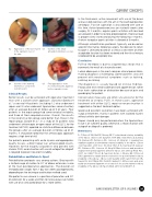
02 Appearance of the bone tunnel 03 in the supinator crest of
the ulna.
04 Insertion of the humeral 05 interference screw in the
isometric point.
Clinical Results
Insertion of the small- articulation interference screw.
In the third week, active movement with use of the brace and assisted exercises with the arm in the overhead position are begun. Passive supination is also allowed with care at this time. Active forced exercises are started 6 weeks after surgery. At 3 months, regular sports activities with low load are allowed in order to restore proprioception. Exercise load is progressively increased during the following 3 months. A return to regular sports activities is allowed at 6 months.
Although rehabilitation protocols typically are based on specific time frames following surgery, the decision to return to sport is ultimately based on a critical assessment of when acceptable function has been restored and the risk of reinjury is believed to be acceptably low.
Conclusion
PLRI of the elbow is due to a ligamentous lesion that is commonly the result of a traumatic event.
Lateral elbow pain is the most common clinical presentation, making diagnosis challenging. Some patients also will present with mechanical symptoms such as locking, catching, or clicking.
The diagnosis is usually based on clinical findings. Provocative tests tend to demonstrate apprehension rather than frank subluxation or dislocation because of pain and guarding by the patient.
Conservative measures are often unsuccessful for the treatment of chronic PLRI. In these cases, operative treatment with either LUCL repair or reconstruction is suggested as the best treatment option.
Good and excellent outcomes have been achieved with surgical treatment, mainly in patients with isolated injuries without arthritic joint damage.
Repair should only be performed when the ligamentous tissue is of sufficient quality; otherwise, a reconstruction with autograft or allograft is preferred.
References
1. O’Driscoll SW, Bell DF, Morrey BF. Posterolateral rotatory instability of the elbow. J Bone Joint Surg Am. 1991 Mar;73(3):440-6. 2. Dunning CE, Zarzour ZD, Patterson SD, Johnson JA, King GJ J. Ligamentous stabilizers against posterolateral rotatory instability of the elbow. J Bone Joint Surg Am. 2001 Dec; 83 A(12):1823-8. 3. Dunning CE, Zarzour ZD, Patterson SD, Johnson JA, King GJ Muscle forces and pronation stabilize the lateral ligament deficient elbow. Clin Orthop Relat Res. 2001 Jul; (388):118-24. 4. Sanchez-Sotelo J, Morrey BF, O’Driscoll SW. Ligamentous repair and reconstruction for posterolateral rotatory instability of the elbow. J Bone Joint Surg Br. 2005 Jan;87(1):54-61.5. Savoie FH 3rd, O’Brien MJ, Field LD, Gurley DJ. Arthroscopic and open radial ulnohumeral ligament reconstruction for posterolateral rotatory instability of the elbow. Clin Sports Med. 2010 Oct;29(4):611-8.
Better results can be achieved with operative treatment than with a conservative approach. Sanchez-Sotelo et al.4 assessed 44 patients (including 12 who underwent repair and 32 who underwent ligamentous reconstruction) after an average duration of follow-up of 6 of years. Four patients in the repair group had some residual instability, and three of them required revision. Overall, the results in the reconstruction group were better than those in the repair group. Savoie et al.5, in a study of 54 patients who underwent arthroscopic or open repair of the LUCL for the treatment of PLRI, reported no statistical difference between the groups after an average duration of follow-up of 41 months. It should be noted that the arthroscopic approach requires a high level of skill.
For patients presenting with acute injuries and appropriate- quality tissues, a direct repair can achieve good results. However, for the majority of patients who present with chronic PLRI, reconstruction with use of autograft or allograft is required to prevent recurrent instability.
Rehabilitation and Return to Sport
Rehabilitation protocols vary among authors. Bracing with a limited range of motion to 30° of elbow extension is one of the main postoperative approaches. The duration of immobilization ranges from 1 day to 6 weeks postoperatively, depending on the technique and fixation method used.
We prefer to use a brace in a position of pronation and 30° of extension for a week and then to increase passive motion with use of an articulated brace for 2 more weeks.
Final appearance of the reconstructed LUCL ligament, before capsular closure.
ISAKOS NEWSLETTER 2019: VOLUME I 39
