
Page 22 - 2020 ISAKOS Newsletter Volume I
P. 22
CURRENT CONCEPTS
Insertional Achilles Tendinopathy Diagnosis and Clinical Presentation
It is widely assumed that insertional Achilles tendinopathy is clinically diagnosed; nevertheless, radiological imaging may be helpful for better defining the clinical findings as well as for preoperative planning if surgery is required. Lateral and axial views may demonstrate calcific tendonitis as well as an abnormal prominence on the posterosuperior side of the posterior tuberosity of the calcaneus.
Both magnetic resonance imaging (MRI) and ultrasound (US) provide additional information to distinguish the different structural abnormalities at the Achilles tendon insertion. Nevertheless, it must be noted that the extreme sensitivity of MRI may also identify structural abnormalities that are not strictly related to clinical symptoms.
Conservative Management
In the acute phase, an initial period of rest or immobilization, along with modified activity, is advisable. Other conservative options include stretching exercises, extracorporeal shock wave therapy, the use of non-steroidal anti-inflammatory drugs, orthotics, and shoe modification; in particular, heel lifts contribute to a consistent reduction of Achilles tendon tension.
Eccentric training, through which the tenon is lengthened during simultaneous muscular contraction, has not demonstrated significant results when used for the treatment of insertional Achilles tendinopathies, although this option has been proven to be effective in cases of non- insertional pain.
Alternatives to eccentric exercises include infiltrations, electrostimulation, and other options aimed to stimulate the healing process in the degenerated tendon.
Surgical Treatment
Patients who do not respond to conservative management may require surgery.
Most clinicians wait at least 3 to 6 months before proceeding with surgery. Surgical procedures include tendon debridement, enthesiophyte resection, gastrocnemius elongation, and posterosuperior calcaneal eminence removal in cases of concomitant pre-insertional symptoms.
In a recent review, two main categories of surgical treatment emerged: debridement alone and debridement combined with tendon augmentation in cases of excessive tendon loss.
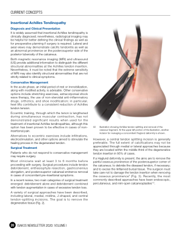
A variety of surgical approaches have been described, including lateral, medial, midline, J-shaped, and central tendon-splitting incisions. The goal is to remove the degenerative tissue (Fig. 2).
02 Illustration showing Achilles tendon splitting and removal of the osseous fragment. At the upper left portion of the illustration, another incision for managing a concomitant Haglund deformity is shown.
However, a central tendon splitting incision is generally preferable. The full extent of calcifications may not be appreciated through medial or lateral approaches because they are located within the middle third of the degenerative tendon insertion in 95% of cases.
If a Haglund deformity is present, the aims are to remove the painful osseous prominence of the posterosuperior corner of the calcaneus; to debride the diseased tendon, if necessary; and to excise the Iinflamed bursal tissue. The surgeon must take care not to damage the tendon insertion when removing the osseous prominence5 (Fig. 3). Recently, the most commonly described approaches have been endoscopic, percutaneous, and mini-open calcaneoplasties1,5.
20 ISAKOS NEWSLETTER 2020: VOLUME I
