
Page 38 - ISAKOS 2020 Newsletter Volume 2
P. 38
CURRENT CONCEPTS
Calipered Kinematic Alignment for Total Knee Arthroplasty
The anterior center of the kinematically aligned baseplate is, on average, closer to the medial border and not the medial one-third of the tibial tubercle, with some variability. The kinematic alignment technique does not refer to mechanical alignment targets such as the mechanical and anatomical axes in the femur and tibia in the coronal plane or the transepicondylar axis and Whiteside’s line in the axial plane.
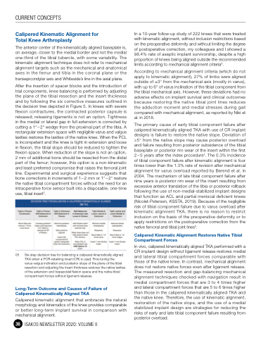
After the insertion of spacer blocks and the introduction of trial components, knee balancing is performed by adjusting the plane of the tibial resection and the insert thickness and by following the six corrective measures outlined in the decision tree depicted in Figure 5. In knees with severe flexion contractures, the contracted posterior capsule is released; releasing ligaments is not an option. Tightness in the medial or lateral gap in full extension is corrected by cutting a 1° – 2° wedge from the proximal part of the tibia. A rectangular extension space with negligible varus and valgus laxities restores the laxities of the native knee. When the PCL is incompetent and the knee is tight in extension and loose in flexion, the tibial slope should be reduced to tighten the flexion space. When reduction of the slope is not an option, 2 mm of additional bone should be resected from the distal part of the femur; however, this option is a non-kinematic and least-preferred compromise that raises the femoral joint line. Experimental and surgical experience suggests that bone corrections in increments of 1–2 mm or 1°–2° restore the native tibial compartment forces without the need for an intraoperative force sensor built into a disposable, one-time use, tibial insert2.
05 Six-step decision tree for balancing a calipered kinematically aligned TKA when a PCR-retaining insert (CR) is used. Fine-tuning the varus-valgus inclination and posterior slope of the plane of the tibial resection and adjusting the insert thickness restores the native laxities of the extension and trapezoidal flexion space and the native tibial compartment forces without ligament releases.
Long-Term Outcome and Causes of Failure of Calipered Kinematically Aligned TKA
Calipered kinematic alignment that embraces the natural morphology and kinematics of the knee provides comparable or better long-term implant survival in comparison with mechanical alignment.
36 ISAKOS NEWSLETTER 2020: VOLUME II
In a 10-year follow-up study of 222 knees that were treated with kinematic alignment, without inclusion restrictions based on the preoperative deformity and without limiting the degree of postoperative correction, my colleagues and I showed a 98.4% rate of aseptic implant survivorship, despite a high proportion of knees being aligned outside the recommended limits according to mechanical alignment criteria3.
According to mechanical alignment criteria (which do not apply to kinematic alignment), 27% of limbs were aligned outside of ±3° from the mechanical axis (mostly in varus), with up to 6° of varus inclination of the tibial component from the tibial mechanical axis. However, these deviations had no adverse effects on implant survival and clinical outcomes because restoring the native tibial joint lines reduces the adduction moment and medial stresses during gait compared with mechanical alignment, as reported by Niki et al. in 2018.
The primary cause of early tibial component failure after calipered kinematically aligned TKA with use of CR implant designs is failure to restore the native slope. Deviation of ≥5° from the native slope may cause posterior overload and failure resulting from posterior subsidence of the tibial baseplate or posterior rim wear of the insert within the first 2 – 5 years after the index procedure4. The 0.3% incidence of tibial component failure after kinematic alignment is four times lower than the 1.3% rate of revision after mechanical alignment for varus overload reported by Berend et al. in 2004. The mechanism of late tibial component failure after nine years is posterior rim wear of the insert resulting from excessive anterior translation of the tibia or posterior rollback following the use of non-medial-stabilized implant designs that function as ACL and partial meniscal deficient knees (Nicolet-Petersen, KSSTA, 2019). Because of the negligible risk of tibial component failure due to varus overload after kinematic alignment TKA, there is no reason to restrict inclusion on the basis of the preoperative deformity or to apply restrictions on the postoperative correction from the native femoral and tibial joint lines3.
Calipered Kinematic Alignment Restores Native Tibial Compartment Forces
In vivo, calipered kinematically aligned TKA performed with a CR implant design without ligament release restores medial and lateral tibial compartment forces comparable with those of the native knee. In contrast, mechanical alignment does not restore native forces even after ligament release. The measured resection and gap-balancing mechanical alignment techniques checked with navigation result in medial compartment forces that are 3 to 4 times higher and lateral compartment forces that are 5 to 6 times higher than those in the calipered kinematically aligned TKA and the native knee. Therefore, the use of kinematic alignment, restoration of the native slope, and the use of a medial stabilized implant design are strategies for reducing the risks of early and late tibial component failure resulting from posterior overload.
