
Page 21 - ISAKOS 2018 Newsletter Volume 2
P. 21
CURRENT CONCEPTS
This entire process is well described in the ISAKOS article on “The Pathogenesis and Classification of Shoulder Stiffness.”1 All of these factors affect the presentation of the patient, the likely natural history, and the outcome of surgical intervention.
Imaging
Radiographs are used to evaluate joint congruency, joint space and articular degeneration, heterotopic ossification, osseous deformity and non-union, and the presence and location of internal fixation.
Computerized tomography (CT) provides a greater resolution of the osseous structures and therefore is very useful for surgical planning. In particular, 2D and 3D CT reconstructions are useful for evaluating mild joint incongruity, degeneration, loose bodies, the morphology of osteophytes (including their size, shape, location, and interaction with the fossae on the anterior and posterior aspects of the elbow), and the proximity of heterotopic ossification to vascular and nerve structures.
Classification
A number of classification systems have been developed for elbow stiffness. We will review some of these systems and highlight their clinical utility.
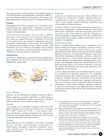
03
Plane of Motion
Stiffness can be classified according to whether it affects flexion-extension, pronation-supination, or both planes of motion. This classification system is practical for evaluating patient impairment and for planning surgery (Fig. 3).
Severity
In this system, the contracture is graded on the basis of the flexion-extension arc of motion as minimal (>91°), moderate (61° to 90°), severe (31° to 60°), or very severe (≤30°). This classification is simple and practical for assessment, prognosis, and surgical decision-making. It can also be used to evaluate impairment and improvement.
Etiology
Elbow stiffness can be classified on the basis of etiology as post-traumatic, micro-traumatic, degenerative or inflammatory, neurogenic, and other (congenital, tumor, burns, infection, etc.). This system is useful for evaluating treatment indications.
Prognosis
Jupiter et al. divided post-traumatic elbow stiffness into 2 categories: simple and complex2. Simple stiffness is characterized by mild to moderate contracture (motion, >80°), no prior surgery, minimal heterotopic ossification, and a well-preserved osseous anatomy.
Complex stiffness is associated with nerve dysfunction, prior nerve transposition, retained hardware, substantial heterotopic ossification, articular incongruity, and an arc of motion of <80°. This system is useful for evaluating the risks and benefits of surgical treatment as the complex form is associated with a higher surgical complication rate and requires more expertise.
Anatomical Location
Morrey classified elbow stiffness into 3 categories on the basis of anatomical location and etiology: extrinsic, intrinsic, and mixed (Morrey, 2005). Extrinsic factors (i.e., extra- articular factors that spare the joint space) include capsular- ligament contracture, muscle-tendon retraction, ulnar nerve neuropathy, heterotopic ossification, and skin contracture. Intrinsic factors (i.e., intra-articular factors) include intra- articular adhesions and deformities, cartilage lesions, loose- bodies, and impinging osteophytes. Mixed factors are those that have both intrinsic and extrinsic elements. Until now, this system has been the most commonly used tool for the classification of joint stiffness.
In 2010, Watts and Bain3 recommended that the capsule be considered as a separate category as it is a separate anatomical structure with a different etiology, treatment, and prognosis. Isolated capsular contractures can be caused by immobilization or hemarthrosis. Capsular contractures also are commonly noted in association with intra-articular pathologies. In cases of elbow stiffness, capsular contracture is often the main problem to be addressed as it is uniquely positioned to interact with both the intra-articular and extra- articular factors.
01 Diagram illustrating the concept of the “elbow machine,” which helps to understanding how the joint moves and highlights the 4 distinct anatomical areas where disorders may affect elbow motion. (Copyright Drs. G. Bain and A. Marinelli).
02 Diagrams illustrating why elbow stiffness is a disabling condition. The blue regions show the areas that are reachable by the hand with a full range of motion of the shoulder and elbow. The red regions represent the much smaller areas that are reachable by the hand with a normal shoulder but a 90° elbow flexion contracture.
03 Diagrams illustrating anatomical factors that can restrict extension
(left) and flexion (right). With restricted extension, it may be necessary to release the contracted anterior soft tissues (anterior capsule, flexor muscles, dermis) and/or to excise any posterior osseous impingement. With restricted flexion, it may be necessary to release the contracted posterior soft tissues (the posterior band of the MCL, posterior capsule, triceps) and/or to excise any anterior osseous impingement. (Copyright Dr. A. Marinelli).
ISAKOS NEWSLETTER 2018: VOLUME II 19
