
Page 37 - ISAKOS 2018 Newsletter Volume 2
P. 37
References
1. Munn J, Sullivan SJ, Schneiders AG. Evidence of sensorimotor deficits in functional ankle instability: a systematic review with meta- analysis. J Sci Med Sport 2010; 13(1): 2-12. 2. Takao M, Innami K, Matsushita T, Uchio Y, Ochi M. Arthroscopic and magnetic resonance image appearance and reconstruction of the anterior talofibular ligament in cases of apparent functional ankle instability. The American journal of sports medicine 2008; 36(8): 1542-7. 3. Vega J, Pena F, Golano P. Minor or occult ankle instability as a cause of anterolateral pain after ankle sprain. Knee surgery, sports traumatology, arthroscopy : official journal of the ESSKA 2016; 24(4): 1116-23. 4. Ferkel RD, Karzel RP, Del Pizzo W, Friedman MJ, Fischer SP. Arthroscopic treatment of anterolateral impingement of the ankle. The American journal of sports medicine 1991; 19(5): 440-6. 5. Ross KA, Murawski CD, Smyth NA, et al. Current concepts review: Arthroscopic treatment of anterior ankle impingement. Foot and ankle surgery : official journal of the European Society of Foot and Ankle Surgeons 2017; 23(1): 1-8.
01 Diagram illustrating the description of anterolateral impingement as proposed by Ferkel et al.4
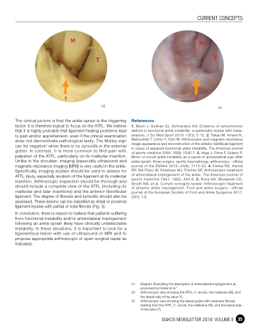
02 Arthroscopic view showing the ATFL (1, arrow), the malleolus (M), and the lateral side of the talus (T).
03 Arthroscopic view showing the lateral gutter with extensive fibrosis starting from the ATFL (1, arrow), the malleolus (M), and the lateral side of the talus (T).
CURRENT CONCEPTS
02
The clinical picture is that the ankle sprain is the triggering factor. It is therefore logical to focus on the ATFL. We believe that it is highly probable that ligament-healing problems lead to pain and/or apprehension, even if the clinical examination does not demonstrate pathological laxity. The Molloy sign can be negative5 when there is no synovitis in the external gutter. In contrast, it is more common to find pain with palpation of the ATFL, particularly on its malleolar insertion. Unlike in the shoulder, imaging (especially ultrasound and magnetic resonance imaging [MRI]) is very useful in the ankle. Specifically, imaging studies should be used to assess for ATFL injury, especially avulsion of the ligament at its malleolar insertion. Arthroscopic inspection should be thorough and should include a complete view of the ATFL (including its malleolar and talar insertions) and the anterior tibiofibular ligament. The degree of fibrosis and synovitis should also be assessed. These lesions can be classified as distal or proximal ligament injuries with partial or total fibrosis (Fig. 3).
In conclusion, there is reason to believe that patients suffering from functional instability and/or anterolateral impingement following an ankle sprain likely have clinically undetectable instability. In these situations, it is important to look for a ligamentous lesion with use of ultrasound or MRI and to propose appropriate arthroscopic or open surgical repair as indicated.
03
ISAKOS NEWSLETTER 2018: VOLUME II 35
