
Page 47 - ISAKOS 2020 Newsletter Volume 2
P. 47
CURRENT CONCEPTS
Metalloproteases seem to stimulate the stem cells from the neural crest which, in a hypoxic environment (e.g. muscle necrosis) leads to pathological bone formations2. Patients affected by transient neuropathies are much more affected by HO than others.
Some patients may have a genetic predisposition to develop HO. Moore-Lotridge et al.3 recently proposed the “two hit mechanism,” whereby HO can result from insufficient protection against nanohydroxyapatite deposits with a failure of macrophage-mediated regression.
Many risk factors have been highlighted in the literature. Some risk factors are recurrent and could change the surgeon’s approach to elbow surgery. Delays (>48 hours) in elbow lesion treatment, prolonged immobilization, and the performance of multiple reduction steps are factors that may be associated with the development of HO. Fracture- dislocation patterns, burns, and central nervous system injuries have been correlated with HO development, but physicians cannot directly change these factors.
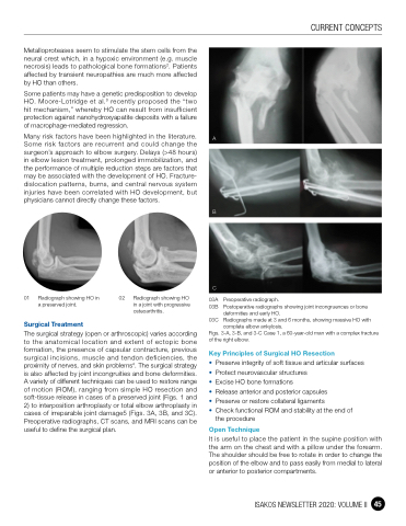
A
B
C
03A Preoperative radiograph.
03B Postoperative radiographs showing joint incongruences or bone
deformities and early HO.
03C Radiographs made at 3 and 6 months, showing massive HO with
complete elbow ankylosis.
Figs. 3-A, 3-B, and 3-C Case 1, a 60-year-old man with a complex fracture of the right elbow.
Key Principles of Surgical HO Resection
• Preserve integrity of soft tissue and articular surfaces • Protect neurovascular structures
• Excise HO bone formations
• Release anterior and posterior capsules
• Preserve or restore collateral ligaments
• Check functional ROM and stability at the end of
the procedure
Open Technique
It is useful to place the patient in the supine position with the arm on the chest and with a pillow under the forearm. The shoulder should be free to rotate in order to change the position of the elbow and to pass easily from medial to lateral or anterior to posterior compartments.
01 Radiograph showing HO in a preserved joint.
Surgical Treatment
02 Radiograph showing HO in a joint with progressive
osteoarthritis.
The surgical strategy (open or arthroscopic) varies according to the anatomical location and extent of ectopic bone formation, the presence of capsular contracture, previous surgical incisions, muscle and tendon deficiencies, the proximity of nerves, and skin problems4. The surgical strategy is also affected by joint incongruities and bone deformities. A variety of different techniques can be used to restore range of motion (ROM), ranging from simple HO resection and soft-tissue release in cases of a preserved joint (Figs. 1 and 2) to interposition arthroplasty or total elbow arthroplasty in cases of irreparable joint damage5 (Figs. 3A, 3B, and 3C). Preoperative radiographs, CT scans, and MRI scans can be useful to define the surgical plan.
ISAKOS NEWSLETTER 2020: VOLUME II 45
