
Page 36 - ISAKOS 2019 Newsletter Vol II
P. 36
CURRENT CONCEPTS
Distal Biceps Endoscopy: All-Endoscopic Exploration, Repair, and Reconstruction of Distal Biceps Tears
Contraindications
Post-traumatic or iatrogenic alteration in the anatomy of the cubital fossa (e.g., as a result of scarring, heterotopic ossification, or vascular or neurological surgery) is a contraindication to the procedure. Similarly, the procedure should be avoided in patients with vascular anomalies and malunited fractures. Surgeon inexperience is a relative contraindication, and familiarity with the anatomical course of major neurovascular structures is necessary in order to prevent any iatrogenic complications.
Surgical Technique
Biceps endoscopy is currently performed with use of 2 different techniques: (1) the single-portal (incision) and endoscopic-assisted technique and (2) the all-endoscopic technique.
Single portal (incision) and endoscopic-assisted technique:
Eames and Bain were the first to describe an anterior portal using a 2.5-cm incision placed 2 cm distal to the elbow crease for endoscopic visualization of the DBT6. The superficial radial nerve and posterior interosseous nerve are at risk of iatrogenic injury with this portal, and retractors used via the same incision significantly decrease the risk of injury to any neurovascular structures4. Phadnis and Bain described the current modification of the endoscopic-assisted footprint repair using a 3 to 5-cm midline longitudinal incision approximately two fingerbreadths distal to the elbow crease1. Endoscopic magnification via the incision permits clear visualization of the anatomy and optimizes debridement and tendon repair. A disadvantage of the single-portal technique is that the viewing and working area is restricted to the length of the incision, which limits its utility for the treatment of retracted tears.
All-endoscopic technique: This technique, described by Bhatia et al., involves the use of separate endoscopic portals for visualization and working2 – 5. The “proximal parabiceps portal” is the main viewing portal and is placed above the elbow crease, and an additional working portal is placed at the level of the tuberosity2. Fluid insufflation is necessary to create a working space, and cannulas are used to protect soft tissues in the forearm. The all-endoscopic technique can be used with suture anchors, interference screws, or cortical buttons5. The technique can be used to repair non- retracted and chronic retracted ruptures, and deficient tendons can be reconstructed endoscopically with use of a graft3. Excellent visualization at every step of the procedure eliminates the need to retract tissues or excessively dissect surrounding soft-tissue planes and thereby prevents associated complications such as heterotopic ossification and neurovascular injury.
Author’s Preferred Technique
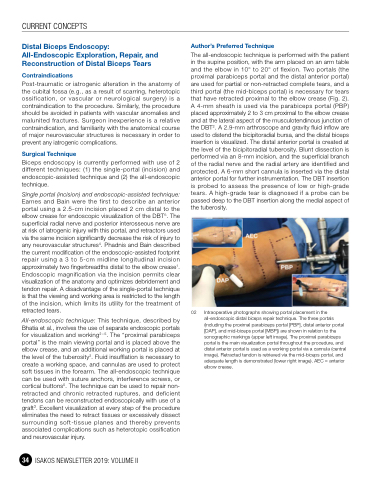
The all-endoscopic technique is performed with the patient in the supine position, with the arm placed on an arm table and the elbow in 10° to 20° of flexion. Two portals (the proximal parabiceps portal and the distal anterior portal) are used for partial or non-retracted complete tears, and a third portal (the mid-biceps portal) is necessary for tears that have retracted proximal to the elbow crease (Fig. 2). A 4-mm sheath is used via the parabiceps portal (PBP) placed approximately 2 to 3 cm proximal to the elbow crease and at the lateral aspect of the musculotendinous junction of the DBT2. A 2.9-mm arthroscope and gravity fluid inflow are used to distend the bicipitoradial bursa, and the distal biceps insertion is visualized. The distal anterior portal is created at the level of the bicipitoradial tuberosity. Blunt dissection is performed via an 8-mm incision, and the superficial branch of the radial nerve and the radial artery are identified and protected. A 6-mm short cannula is inserted via the distal anterior portal for further instrumentation. The DBT insertion is probed to assess the presence of low or high-grade tears. A high-grade tear is diagnosed if a probe can be passed deep to the DBT insertion along the medial aspect of the tuberosity.
02 Intraoperative photographs showing portal placement in the all-endoscopic distal biceps repair technique. The three portals (including the proximal parabiceps portal [PBP], distal anterior portal [DAP], and mid-biceps portal [MBP]) are shown in relation to the sonographic markings (upper left image). The proximal parabiceps portal is the main visualization portal throughout the procedure, and distal anterior portal is used as a working portal via a cannula (central image). Retracted tendon is retrieved via the mid-biceps portal, and adequate length is demonstrated (lower right image). AEC = anterior elbow crease.
34 ISAKOS NEWSLETTER 2019: VOLUME II
