
Page 38 - ISAKOS 2019 Newsletter Vol II
P. 38
CURRENT CONCEPTS
Distal Biceps Endoscopy: All-Endoscopic Exploration, Repair, and Reconstruction of Distal Biceps Tears
Key Points and Technical Tips
• Preoperative sonography is performed, and the course of major neurovascular structures, the bicipital tuberosity, and the retracted tendon end are marked3.
• The elbow joint is flexed 10° to 20° for initial portal placement, and a support under the distal part of the humerus is used intraoperatively to achieve extension when necessary.
• The “proximal parabiceps portal” is angulated dorsally and toward the sonographically marked tuberosity, and the sheath is advanced approximately 7 cm. The sheath should pass in a smooth passage until the bare tuberosity area is felt2.
• The proximal parabiceps portal is adjacent to the lateral cutaneous nerve, and the radial artery is at significant risk of injury when the distal anterior portal is used. The risk is minimized by placing the proximal parabiceps portal just below the musculotendinous junction and in close apposition to the DBT. The distal anterior portal should be placed with open dissection via an 8-mm incision, and the superficial radial nerve and radial artery should be identified and protected.
• The adequacy of tendon length (which usually measures 7 to 8 cm) is judged by the ability to pull the DBT externally to the distal anterior portal3.
• The shuttle suture for the passage of the DBT from mid- biceps portal to distal anterior portal follows the track of the proximal parabiceps portal sheath. The suture is passed through this sheath and then is withdrawn into mid-biceps portal. This provides a safe passage of the DBT under the brachial bifurcation and into the distal anterior portal3.
• Failure of the sutures to slide within the implants results in suboptimal tendon-bone contact area. The consequent gap formation may result in suboptimal healing and may predispose the DBT to rerupture. Gap formation is possible with any fixation device; however, the dual-anchor technique has shown consistent and optimal tendon-bone contact area5.
• The reattachment site on the tuberosity is important. Footprint repair results in better wrapping of the tendon around the medial tuberosity, and probably improves terminal supination strength1. However, the proximal radioulnar space reduces significantly from the supinated to the pronated position, and this reduction is most evident in the distal aspect of the tuberosity. Postoperative DBT impingement in the radioulnar space may be prevented by avoiding techniques that increase the thickness of the tendon and by using a reattachment site at the proximal aspect of the tuberosity7.
36 ISAKOS NEWSLETTER 2019: VOLUME II
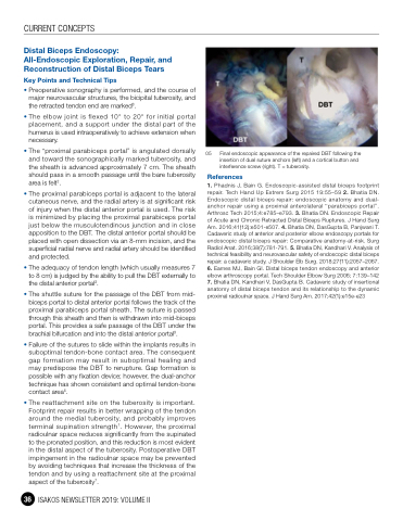
05 Final endoscopic appearance of the repaired DBT following the insertion of dual suture anchors (left) and a cortical button and interference screw (right). T = tuberosity.
References
1. Phadnis J, Bain G. Endoscopic-assisted distal biceps footprint repair. Tech Hand Up Extrem Surg 2015 19:55–59 2. Bhatia DN. Endoscopic distal biceps repair: endoscopic anatomy and dual- anchor repair using a proximal anterolateral ‘‘parabiceps portal’’. Arthrosc Tech 2015;4:e785–e793. 3. Bhatia DN. Endoscopic Repair of Acute and Chronic Retracted Distal Biceps Ruptures. J Hand Surg Am. 2016;41(12):e501-e507. 4. Bhatia DN, DasGupta B, Panjwani T. Cadaveric study of anterior and posterior elbow endoscopy portals for endoscopic distal biceps repair: Comparative anatomy-at-risk. Surg Radiol Anat. 2016;38(7):781-791. 5. Bhatia DN, Kandhari V. Analysis of technical feasibility and neurovascular safety of endoscopic distal biceps repair: a cadaveric study. J Shoulder Elb Surg. 2018;27(11):2057–2067. 6. Eames MJ, Bain GI. Distal biceps tendon endoscopy and anterior elbow arthroscopy portal. Tech Shoulder Elbow Surg 2006; 7:139–142 7. Bhatia DN, Kandhari V, DasGupta B. Cadaveric study of insertional anatomy of distal biceps tendon and its relationship to the dynamic proximal radioulnar space. J Hand Surg Am. 2017;42(1):e15e-e23
