
Page 30 - ISAKOS 2018 Newsletter Volume 2
P. 30
CURRENT CONCEPTS
Update on Meniscal Allograft Transplantation
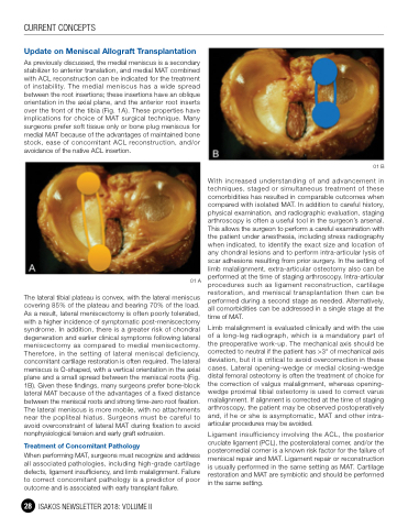
As previously discussed, the medial meniscus is a secondary stabilizer to anterior translation, and medial MAT combined with ACL reconstruction can be indicated for the treatment of instability. The medial meniscus has a wide spread between the root insertions; these insertions have an oblique orientation in the axial plane, and the anterior root inserts over the front of the tibia (Fig. 1A). These properties have implications for choice of MAT surgical technique. Many surgeons prefer soft tissue only or bone plug meniscus for medial MAT because of the advantages of maintained bone stock, ease of concomitant ACL reconstruction, and/or avoidance of the native ACL insertion.
01 A
The lateral tibial plateau is convex, with the lateral meniscus covering 85% of the plateau and bearing 70% of the load. As a result, lateral meniscectomy is often poorly tolerated, with a higher incidence of symptomatic post-meniscectomy syndrome. In addition, there is a greater risk of chondral degeneration and earlier clinical symptoms following lateral meniscectomy as compared to medial meniscectomy. Therefore, in the setting of lateral meniscal deficiency, concomitant cartilage restoration is often required. The lateral meniscus is O-shaped, with a vertical orientation in the axial plane and a small spread between the meniscal roots (Fig. 1B). Given these findings, many surgeons prefer bone-block lateral MAT because of the advantages of a fixed distance between the meniscal roots and strong time-zero root fixation. The lateral meniscus is more mobile, with no attachments near the popliteal hiatus. Surgeons must be careful to avoid overconstraint of lateral MAT during fixation to avoid nonphysiological tension and early graft extrusion.
Treatment of Concomitant Pathology
When performing MAT, surgeons must recognize and address all associated pathologies, including high-grade cartilage defects, ligament insufficiency, and limb malalignment. Failure to correct concomitant pathology is a predictor of poor outcome and is associated with early transplant failure.
28 ISAKOS NEWSLETTER 2018: VOLUME II
01 B
With increased understanding of and advancement in techniques, staged or simultaneous treatment of these comorbidities has resulted in comparable outcomes when compared with isolated MAT. In addition to careful history, physical examination, and radiographic evaluation, staging arthroscopy is often a useful tool in the surgeon’s arsenal. This allows the surgeon to perform a careful examination with the patient under anesthesia, including stress radiography when indicated, to identify the exact size and location of any chondral lesions and to perform intra-articular lysis of scar adhesions resulting from prior surgery. In the setting of limb malalignment, extra-articular osteotomy also can be performed at the time of staging arthroscopy. Intra-articular procedures such as ligament reconstruction, cartilage restoration, and meniscal transplantation then can be performed during a second stage as needed. Alternatively, all comorbidities can be addressed in a single stage at the time of MAT.
Limb malalignment is evaluated clinically and with the use of a long-leg radiograph, which is a mandatory part of the preoperative work-up. The mechanical axis should be corrected to neutral if the patient has >3° of mechanical axis deviation, but it is critical to avoid overcorrection in these cases. Lateral opening-wedge or medial closing-wedge distal femoral osteotomy is often the treatment of choice for the correction of valgus malalignment, whereas opening- wedge proximal tibial osteotomy is used to correct varus malalignment. If alignment is corrected at the time of staging arthroscopy, the patient may be observed postoperatively and, if he or she is asymptomatic, MAT and other intra- articular procedures may be avoided.
Ligament insufficiency involving the ACL, the posterior cruciate ligament (PCL), the posterolateral corner, and/or the posteromedial corner is a known risk factor for the failure of meniscal repair and MAT. Ligament repair or reconstruction is usually performed in the same setting as MAT. Cartilage restoration and MAT are symbiotic and should be performed in the same setting.
