
Page 40 - ISAKOS 2018 Newsletter Volume 2
P. 40
CURRENT CONCEPTS
ACL Laxity Evaluation with Slice Imaging: Applicability of the Porto-Knee Testing Device
References
1. Tashman, S., Collon, D., Anderson, K., Kolowich, P., & Anderst, W. (2004). Abnormal rotatory knee motion during running after anterior cruciate ligament reconstruction. Am J Sports Med, 32(4), 975-983. 2. Espregueira-Mendes, J., Pereira, H., Sevivas, N., Passos, C., Vasconcelos, J. C., Monteiro, A., Oliveira, J. M., & Reis, R. L. (2012). Assessment of rotatory laxity in anterior cruciate ligament-deficient knees using magnetic resonance imaging with Porto-knee testing device. Knee Surg Sports Traumatol Arthrosc, 20(4), 671-678. 3. Espregueira- Mendes, J., Andrade, R., Leal, A., Pereira, H., Skaf, A., Rodrigues- Gomes, S., Oliveira, J. M., Reis, R. L. & Pereira, R. (2017). Global rotation has high sensitivity in ACL lesions within stress MRI. Knee Surg Sports Traumatol Arthrosc, 25(10), 2993-3003. 4. Tashiro Y, Okazaki K, Miura H, Matsuda S, Yasunaga T, Hashizume M, Nakanishi Y, Iwamoto Y (2009) Quantitative assessment of rotatory instability after anterior cruciate ligament reconstruction. Am J Sports Med 37:909–916
03
04
Take-Home Message
Instrumented laxity measurements made with use of MRI or CT scans during the application of stress with use of the PKTD provide a clear picture of both the structural and functional status of the knee. Thus, a knee with a structural lesion may be found to be functionally competent, which may support a conservative approach. Alternatively, a knee with a structurally continuous ligament may be found to be non- functional (or even to exhibit a chewing-gum effect), which may require surgery.
04
05
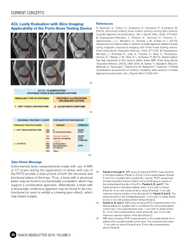
Panels A through E: MRI scans showing the PKTD measurements in the lateral plateau (Panels A, B and C) and medial plateau (Panels D and F) for a patient with a partial ACL rupture. PKTD assessment showed important internal rotation (+9 mm) following a rupture of
the posterolateral bundle of the ACL. Panels A, B, and C: The measurements in the lateral plateau were -2 mm with no stress (Panel A), 6 mm with posteroanterior stress (Panel B), 7 mm with maximum internal rotation of the tibia (panel C). Panels D and E: The measurements in the medialplateauwere -4 mm with no stress (Panel D) and 3 mm with posteroanterior stress (Panel E).
Panels A, B, and C: MRI scans showing PKTD measurements in the lateral plateau for a patient with a combined ACL and posterolateral corner injury. The measurements were -1 mm with no stress (Panel A), 14 mm with posteroanterior stress (Panel B), and -6 mm with maximum external rotation of the tibia (Panel C).
MRI scans showing PKTD measurements in the medial plateau for a patient with a posteromedial corner injury. The measurements were
-2 mm with no stress (Panel A) and -6 mm with posteroanterior stress (Panel B).
05
03
38 ISAKOS NEWSLETTER 2018: VOLUME II
