Page 15 - Layout 1
P. 15
LIFESTYLE
Surgical technique (deltopectoral approach)
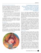
“Outstanding instructor and
After combined general and interscalene regional anaesthesia arthroscopist. He teaches his fellows
the patient is positioned in the beach chair position with
to open their minds to a thorough
approximately 45 degrees of tilt of the backrest; a 10 cm skin
incision is made, approximately 2-3cm lateral and parallel understanding of the shoulder,
to the deltopectoral interval, beginning at the level of the
coracoid and extending distally towards the mid-humerus.
providing for endless possibilities”
A more lateralized approach to classic deltopectoral skin –Tom Christensen
incision offers the following advantages:
1. It ensures that the Cephalic vein will always be found The arm is then flexed to approximately 60 degrees and
externally rotated approximately 20 degrees. The plane
medial to our skin incision and so can be quickly identified.
between the subscapularis tendon and the joint capsule is
2. A lateralized incision improves visualisation of the glenoid.
developed and the tendon is retracted superiorly, exposing
3. It creates an overlap of normal skin over the deltopectoral
the antero-inferior joint capsule. The axilliary nerve is located
interval ensuring natural tissue planes to be restored post and exposed adjacent to the joint capsule inferior to the
operatively.
glenoid neck and a small Hohmann retractor is placed to
A standardised dissection of the deltopectoral interval is protect the nerve.
performed till the subscapularis is reached. The superior and The inferior arthrotomy is then performed into the axillary pouch.
inferior borders of the subscapularis tendon are identified, The capsulotomy is extended laterally along the capsular
using the anterior circumflex humeral vessels as a guide to attachment to the humerus and medially along the glenoid
the inferior aspect of the tendon.
neck, and the capsule is fully excised. Once completed, a
second retractor is placed at the inferior aspect of the humeral
Inferior Window
head exposing the osteophytes anteriorly and inferiorly. The
The inferior window is the first arthrotomy made and is
osteophytes are resected using curved osteotomes and
used to expose the inferior aspect of the joint for removal of rongeurs. Complete removal of the osteophytes can be
osteophytes. The inferior window can also be used to check
confirmed by palpation. It is important to ensure that the
component positioning during trialling and insertion. The posterior osteophytes, which are difficult to access, have
window is opened below the subscapularis tendon, medially been removed. After removal of the osteophytes, the inferior
as far as the glenoid and laterally to the tendon insertion of aspect of the glenoid can be visualised and an inferior soft
subscapularis.
tissue release performed. The position of the axillary nerve
To open the window, a 1cm partial tenotomy of the superior should be considered at all times during the capsulotomy and
part of the pectoralis major tendon is performed, exposing removal of the osteophytes.
the underlying latissimus dorsi tendon. The anterior circumflex
humeral vessels are ligated and cauterized.
Superior Window
Attention is then turned to the superior window. This window
opens the joint via the rotator interval and is used for further
joint preparation, instrumentation, trialling and insertion of the
prosthesis. To improve visualisation, the shoulder is extended
approximately 40 degrees in relation to the patient’s body and
in neutral rotation. The rotator interval tissue is completely
excised including the coracohumeral ligament, superior
glenohumeral ligament, proximal portion of the biceps tendon
and joint capsule. The rotator interval is trapezoidal in shape
and extends from the anterior border of the supraspinatus
tendon to the glenoid medially and then laterally along the
superior border of the subscapularis tendon. The lateral
border is formed by the bicipital groove.
To open the interval the tissue is incised along the superior
border of the subscapularis tendon. The biceps tendon can
then be identified intraarticularly and the bicipital groove
can opened. In cases where the superior border of the
subscapularis tendon and CHL is not easy to identify, the
bicipital groove at the upper part of the epiphysis will guide the
surgeon to the rotator interval. A biceps soft tissue tenodesis
is performed within the groove and the proximal portion of
the biceps tendon is excised, along with all remaining interval
tissue.
04
ISAKOS NEWSLETTER 2014: Volume II 13