Page 13 - Layout 1
P. 13
LIFESTYLE
5. When intra-articular visualization is adequate, the greater
tuberosity is prepared, and a double-loaded suture anchor
4.5 or 5.5 mm is placed. With small tears, the anchor is
placed just posterior to the biceps groove; in medium-
sized tears, the anchor is placed more posteriorly. If no
cuff tear is evident, the anchor is placed in the groove.
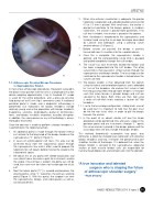
6. Next, the biceps is tenodesed to the supraspinatus and
humeral head using the lasso-loop technique described
by Lafosse and colleagues using a piercing suture
retriever device (1) (Figure 2).
7. Before sutures are passed, the biceps is partially
transected near its insertion into the superior labrum.
8. Once this is complete, the supraspinatus tendon is
pierced, and the corresponding suture limb is grasped
and pulled completely through the cuff tendon.
9. The suture has now essentially locked the biceps and
has been incorporated into the cuff tendon. The other
suture limb is then passed in similar fashion through the
supraspinatus and biceps tendon. The lasso loop can be
02
switched so the supraspinatus tendon is locked when the
sutures are tied.
10. The biceps tendon is than detached from the labrum 1.1. Arthroscopic Proximal Biceps Tenodesis
using the lateral and anterolateral portals and is debrided to Supraspinatus Tendon
to 1 cm of the tenodesis site and our first suture is tied;
In most of our arthroscopic procedures, the patient is placed in
the beach-chair position with the arm in a longitudinal traction the limb passing freely through both tendons is the post
such that when tension is placed, biceps and cuff are
device weighing approximately 3 kg. A standard 30° scope
is used for the entire procedure, and a pressure-sensitive secured to the bone. Both limbs are then tied using
fluid pump set to 60 mm Hg is commonly used. A traditional a series of half-hitch knots creating a “pulver taft”-like
tenodesis.
posterior portal is made, and a diagnostic arthroscopy is
performed. Our indications for biceps tenodesis include 11. Due to the lasso-loop configuration, sliding knots cannot
relatively young and active population with biceps tendonitis be used; but it is important to note that the post must
(hyperemia, synovitis), tendinopathy, partial or full-thickness be the the non lasso stitch; in order to pull the biceps
tears, and biceps instability secondary to pulley disruption; towards the anchor.
in addition, the subscapularis or any cuff pathology is always 12. In the event of an absent rotator cuff tear the whole
thoroughly examined.
procedure will be performed intra articularly, scope in the
posterior portal and our instruments through “D” portal;
Once the decision is made to perform a biceps tenodesis, it
is performed in the following fashion:
the suture anchor is placed in the biceps groove and the
lasso loop stitch will only include the biceps tendon.
1. An additional portal is made through the rotator interval
This method theoretically accomplish two goals: one,
just anterior to the long head of the biceps tendon or the
supraspinatus (“D” portal in Figure 1).
eliminates a potential shoulder pain generator with proximal
biceps tenodesis and two, it restores the glenohumeral joint
2. In cases of anterosuperior cuff tears, this area can be stabilizer function of the biceps. It is believed that, when the
accessed easily from the subacromial space through
biceps tendon is secured to the supraspinatus, opposing
the supraspinatus tear and is often used to prepare the vectors of both muscle tendon units are responsible for
tuberosity for cuff repairs before moving the scope to the
humeral head depression and compression into the glenoid.
subacromial space.
3. We obtain this or any portal with needle localization and
use a blunt trocar to create a path for instruments and/or
the scope. If no cuff tear is evident, the portal can still be
“A true innovator and talented
used, but care must be taken not to damage the intact
cuff.
surgeon who is shaping the future
4. Next the lateral portal (“C”) is created and becomes the of arthroscopic shoulder surgery”
viewing portal, while “D” becomes the primary working
–Ruth Delaney
portal (Figure 1). With this portal, the surgeon can view
the supraspinatus, biceps, and subscapularis as well as
anchor placement.
ISAKOS NEWSLETTER 2014: Volume II 11