Page 31 - Layout 1
P. 31
CURRENT CONCEPTS
Patella Dislocations
(Isolated) Patellofemoral Arthritis
Historically, PF instability was felt to be primarily a disorder of PF arthritis has been a relatively undiagnosed as a cause of
females. (Figure 1) However, a systematic review of primary knee pain. (Figure 3) Though studies looking at sex as a risk
traumatic patella dislocations reveals a nearly equal female for isolate PF arthritis are sparse, the available clinical studies
to male distribution. Of 22 articles reviewed, the total number show an overwhelmingly female preponderance. McAlindon
of first-time PF dislocators was 1765, with a male / female et al. reviewed radiographs of 273 subjects with complaints
ratio of 46% / 54%, average age 21.5 years. A second study of knee pain; isolated PF arthritis was present more than twice
looking at the epidemiology and natural history of acute as often in females (24%) vs. males (11%). The incidence of
patella dislocations suggests a similar incidence of males and combined medial and PF compartment arthritis showed equal
females in primary patella dislocations, with a preponderance incidence between males (7%) and females (6%). Iwano et al.
of recurrent dislocations occurring in females. In this study, and colleagues reviewed a series of 108 knees in 69 patients
risk was highest among females age 10–17 years.
with PF arthritis; 93% were female. In a large French multi-
center review of 578 patients with isolated PF arthritis, 72%
A report of 127 patients with primary dislocations at two of the patients were female.
Finnish trauma centers followed for 7 years revealed a slight
female preponderance (64%). In analyzing risk factors, the
Conclusion
authors found two risk factors for recurrent instability were
initial contralateral instability and young age. Females with Review of the clinical literature to date suggests sexual
dimorphism in the presentation of PF disorders, with an
open tibial epiphysis at the time of the initial dislocation had overrepresentation of these disorders in females. However, to
the worst prognosis for recurrent instability.
date there has not been shown a reason to alter our treatments
A seminal study from Lyon, France analyzed factors of patella of these disorders based on the patient’s sex. Continued
instability using standardized imaging to identify factors related study of PF disorders and their treatments should include sex
to patella instability. In their review of 110 patients, 83 were as a variable, in hope of providing better prevention, treatment
female (75%). In reviewing risk factors for patella instability, and care of these disorders.
the authors found that trochlear dysplasia, as defined by the
crossing sign, was present in 96% of patients with objective
patella instability. (Figure 2)
A recent study analyzed imaging of patella instability patients
compared to a control group. The goal was to identify
sex-related differences in the anatomy of lateral patellar
dislocations. The authors found that trochlear dysplasia and
the TT-TG distance is more pronounced in women who
experience patellar dislocation.
A cautious assumption of the current literature suggests that
females are more likely to suffer recurrent patella dislocations
than their male counterparts. Reasons for this are likely multi-
factorial; including anatomic and neuromuscular factors. We
know that compared with males, females display lower knee
flexion angles with activities, with greater knee valgus angles
and quadriceps activation. There is a higher prevalence of
dysplastic distal femora among females.
Understanding sexual dimorphism in neuromuscular and
anatomic risk factors is key. The clinician should be especially
vigilant with their female patients when discussing re-injury
risk, inclusive of known anatomic factors of instability, and be
aware of potential neuromuscular factors during rehabilitation 03
back to sporting activities after a patella dislocation.
01 Fig 1 Axial MR image of an acute patella dislocation depicting classic
bone bruising, torn MPFL, and large effusion
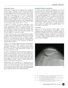
02 Fig 2 True lateral Radiograph depicting Type B Trochlea Dysplasia with
Crossing sign and supratrochlear bump
03 Fig 3 An axial radiograph showing loss of lateral patallofemoral joint
space indicative of patellofemoral arthritis
ISAKOS NEWSLETTER 2013: Volume II 29