Page 33 - Layout 1
P. 33
CURRENT CONCEPTS
Formal postoperative rehabilitation commenced at 2 weeks
with institution of gentle passive range of motion (ROM) limited
to 90 degrees of flexion and abduction with “subscapularis
precautions” (no active internal rotation or passive external
rotation beyond neutral) x 6 weeks. Sling immobilization was
discontinued at 6 weeks with commencement of progressive
active ROM and strengthening thereafter.
02 03
05a
04
Plain film imaging was obtained (Figure 1). MRI demonstrated
a recurrent Bankart lesion with further capsulolabral injury
consistent with an anterior labral periosteal sleeve avulsion
(ALPSA) as well as insufficiency and near complete fatty
degeneration of the subscapularis.(Figure 2,3) By both CT
and MRI anterior glenoid bone loss was calculated to be 22%
using the circle method.(Figure 4)
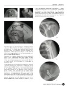
A decision was made to proceed with revision stabilization
utilizing a fresh oseochondral distal tibia allograft for bony
glenoid reconstruction which was fixated utilizing 2 parallel
05b
3.5mm fully threaded cortical screws with a washer (Synthes
Inc, West Chester, PA, USA).(Figure 5) as described by
Provencher, et al.
As the subscapularis was not able to be mobilized or repaired,
a pectoralis transfer was performed. A modification of the
technique described by Resch, et al was performed based
on the anatomic study of Fung, et al where the anterior
and posterior leaflets of the pectoralis major attachment
to the humerus were separated and the anterior leaflet
(including the clavicular head and upper 3 to 5 sternal head
attachments) was mobilized and transferred.(Figure 6) The
split pectoralis was then passed subcoracoid, anterior to the
musculocutaneous nerve and posterior to the conjoint tendon
and was secured to the lesser tuberosity with two 4.5mm
double-loaded suture anchors (Arthrex Inc, Naples, FL, USA).
05c
ISAKOS NEWSLETTER 2013: Volume II 31