Page 35 - Layout 1
P. 35
CURRENT CONCEPTS
Additionally, the decision to utilize a free graft for glenoid
reconstruction was complimented by the decision that a
pectoralis transfer would likely be required for treatment of this
irreparable subscapularis tear. It has been well demonstrated
that pectoralis major transfers perform superiorly when
transferred in a subcoracoid fashion, more closely mirroring
the vector of the native pull of the subscapularis it is being
utilized to compensate for. Thus, it was felt to be preferable
to not perform Latarjet reconstruction, in order to preserve
the conjoint tendon and coracoid to take advantage of this
biomechanical principle. Furthermore, based on the anatomic
study performed by Fung, et al demonstrating that the 08a 08b
pectoralis major is indeed made up of anterior and posterior
leaflets without a rotational component to the terminal tendon
insertion, the techniques described by both Resch and Gerber
was modified. This modification allows transfer of the anterior
leaflet of the pectoralis major tendon, encompassing the
clavicular and upper 3 to 5 sternal attachments, preserving
the remaining posterior leaflet, which in this thin female was
felt to be beneficial for primarily cosmetic purposes of her
upper chest.
Though failure of open treatment for shoulder instability with
glenoid bone loss or subscapularis insufficiency has been
well reported, to our knowledge, this particular constellation
of findings has not been reported together previously. What 08c
makes this patient situation unique is the specific set of
conditions that led to ultimate decision making regarding the
use of free graft for glenoid reconstruction and pectoralis major
tendon transfer with the modification of previously described
techniques as stated above. This case demonstrates a
successful and novel treatment strategy in this complicated
recurrent instability patient that may be useful to surgeons
treating this particular set of reasons for failure of open repair
in recurrent anterior shoulder instability.
01 Fig 1
AP (a), Scapular Y (b) and Axillary views of the left shoulder were
obtained which demonstrated blurring of the anterior glenoid
margin (arrow)
02 Fig 2
Axial MRI demonstrating capsulolabral tear and irreparable
subscapularis insufficiency
03 Fig 3
Sagital MRI demonstrating near complete fatty degeneration of
the subscapularis – indicating an irreparable rotator cuff tear.
04 Fig 4
Sagital Image demonstrating 22% glenoid bone loss as
calculated by the “circle method” – beyond what is felt to be a
“critical “ sized glenoid defect.
05 Fig 5
Postoperative AP, Scapular Y and Axillary views with distal tibia
allograft in place.
06 Fig 6
Intraoperative image demonstrating separation of the anterior
and posterior leaflets of pectoralis major tendon (a) and
demonstration of pectoralis transfer passed subcoracoid prior to
fixation to the lesser tuberosity (b)
07 Fig 7
Axial CT arthrogram demonstrating anatomic glenoid
reconstruction with healed allograft and excellent integrity of the
pectoralis transfer (a) and Sagital CT arthrogram demonstrating
glenoid reconstruction
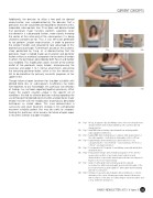
08 Fig 8
Clinial post-operative photographs demonstrating no cosmetic
deformity of the chest following pectoralis transfer, full motion
with resolution of her prior recognized hyper-external rotation and
a now symmetric belly-press.
ISAKOS NEWSLETTER 2013: Volume II 33