Page 36 - Layout 1
P. 36
WORST CASE SCENARIO
Air Embolus During Clavicle The patient was intubated. A chest tube was inserted but no
Internal Fixation with a Plate
pneumothorax was identified. A vascular surgeon aspirated air
from the right atrium via a central line. The patient deteriorated
and was declared deceased.
The coroners report identified;
– Air in the right atrium
– A 25mm perforation of the subclavian vein directly below
Gregory Bain, MBBS, FRACS, the most medial screw hole.
FA(Ortho)A, PhD1,2
Reported cause of death: “air embolism and severe
haemorrhage”.
Discussion
The risk of penetration and subsequent air emboli depends
on the:
Kevin Eng (FRACS)1,2 1 Anatomy of the subclavian vein and artery
Matthias A Zumstein, MD3
2 Clavicle dimensions
1 Department of Orthopedics and Traumatology, University 3 Surgical instruments and technique
of Adelaide, South Australia, AUSTRALIA
2 Department of Orthopaedics and Trauma, Modbury Anatomy Of Subclavian Vein And Artery
The subclavian vessels begin posterosuperiorly and pass
Public Hospital and Royal Adelaide Hospital, South
Australia, AUSTRALIA
inferior to the clavicle at the lateral end. (Figure 1–3) The vein
3 Shoulder and Elbow Unit, Dept. Orthopaedic Surgery lies anterior to the artery, closer to the posterior border of the
Traumatology, University of Bern, Inselspital, Bern, clavicle. The subclavian vein is only 5 mm behind the clavicle
SWITZERLAND
in its medial third, and may even be adherent to the clavicle,
particularly if the anatomy is distorted such as in cases of
There was a fatality in Brisbane, Queensland during internal revision surgery, infection or non union.
fixation of a clavicle fracture, which was reviewed in the
Queensland Coroner’s Court. At the request of Dr. Phil Duke,
then President of the Shoulder and Elbow Society of Australia,
we reviewed the coroner’s records, reviewed the literature,
and published an article in JBJS in 2013.
This article provides a brief discussion of this devastating
case.
We provide this important information in the hope that it may
help prevent a recurrence of this unfortunate event.
Introduction
01
Clavicle fractures are common, and there is a trend towards
internal fixation, especially if there is shortening of > 2cm.
Reported major complications include, subclavian vessel
thrombosis, arterial injury, pseudoaneurysm and neurological
injury.
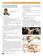
Case Report
A 34yo man sustained an isolated midshaft clavicle fracture,
which was managed with ORIF one month following the
fracture. The patient was positioned supine with a sandbag
under the affected shoulder. A laryngeal mask was used. The 02
clavicle was fixed with a 6 hole locking distal lateral clavicle
plate positioned superiorly. A Bristow elevator was placed
on the inferior surface of the clavicle whilst drilling. Locking
screws with a locking guide were utilised. The final screw
was the most medial. On withdrawal of the drill profuse low
pressure bleeding was noted.
The plate was removed and the bleeding subclavian vein
controlled. However, the patient went into shock despite
hemorrhage control and fluid resuscitation.
34 ISAKOS NEWSLETTER 2013: Volume II
03