Page 37 - Layout 1
P. 37
WORST CASE SCENARIO
The characteristics of venous and arterial bleeding are Trajectory:
different. Venous bleeding is continuous, dark and lower The drill and screws should be aimed away from the subclavian
pressure. It may be difficult to control, and instrumentation vessels where practical. A superior plate (screw trajectory
of the vein will tend to tear the wall. Arterial bleeding is high superior to inferior) is safest medially. An anterior plate may be
pressure and pulsatile. It can be clamped and repaired. best for the middle and lateral thirds where the vein is inferior.
Venous bleeding is a more life threatening and more difficult New generation plates are likely to be designed with a “twist, ”
to manage.
which would allow screws to be correctly directed to minimise
Clavicle Dimensions
risk of vascular injury.. Moulded pelvic reconstruction plates
have previously been used in this fashion.
The clavicle dimensions are highly variable. The smallest
diameter may be as little as 6.7mm in the mid diaphysis. If the
subclavian vein is adherent to the clavicle, a drill penetration
of just over 7 mm may damage the vein.
05
04a
Alternative devices:
No vascular injuries have been reported with intramedullary
nailing. A non union would alter the canal and make passage
of a nail difficult.
04b
Pre-Operative Assessment
Pre-operative assessment is important in all surgery. Complex
cases, such as those with extensive comminution, previous
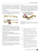
surgery, tumors or extensive osteoporosis may be at higher 01 Fig 1 CT Angiogram: The right SCV (white vessel) is seen passing
risk of vascular injury. Assessment of the plain radiographs
should be performed in all cases. CT angiography in complex directly below the second Quarter of the clavicle. The left
subclavian artery is also marked.
cases will assist in understanding the details of the fracture 02 Fig 2
Superior view of left clavicle, dimensions and vessels:
and vascular structures.
Median width and length of the dry bone clavicles are shown
(mm). The position of the axillary vein (V) and artery (A) are
Surgical Instruments And Technique
represented as a ratio of clavicle length. 0.33 is 1/3 of length
Drill bits and screws may damage the vessels either acutely measured from sternum. S, Sternal end; AC, acromial end. At
risk area is the 2nd quarter. Modified from Galley, Watt, Bain
or by chronic irritation. The risk of drill penetration depends on
depth and trajectory.
JSES 2009.
03 Fig 3
The dangerous trajectory in the middle of the clavicle. Note the
Depth control:
vessels lie posterior to the clavicle medially and inferior to the
Drill stops are becoming more common, however, the authors clavicle laterally [8]. Both lateral and medial the vein is closer to
strongly caution on the use of drill stops as this can lead to the clavicle than the artery. (From Sinha JBJS Am 2011)
04a Fig 4a Anterior view of left clavicle and vessels:
an assumption of 100% safety. The depth of the clavicle is 4a Height of clavicle (mm) at various positions. Note
highly variable, the surgeon may drill obliquely across the
considerable variability. Narrowest clavicle was 7mm.
bone, and not be at its widest position. If the vein is adherent 04b Fig 4b
Anterior view of left clavicle and vessels:
to the clavicle, then it could still be injured. The surgeon must 4b The minimum distance from the superior clavicle to the
superior aspect of the axillary artery (A) was 22 mm. However,
not under any circumstance assume that the stop will ensure
safety!
the vein lies closer. NOTE: the vein can lie on the posterior
periostium of the intact clavicle. A, axillary artery; S, Sternal
Blunt retractors are used but require more dissection, and in end; CC, costocoracoid membrane; SM, subclavius muscle; V,
this case were not effective. Unicortical screw fixation would axillary vein; AC, acromial end. Modified from Galley, Watt, Bain
virtually eliminate risk of vessel penetration but may lead to JSES 2009.
construct failure from screw “pull-out”.
05 Fig 5
Concept of the curved wave plate that directs the screws
away from the vessels. Medial screws from superior to inferior,
Middle screws are unicortical, and lateral screws from anterior
to posterior. (Concept has been presented in open meeting,
therefore not patent protected)
ISAKOS NEWSLETTER 2013: Volume II 35