Page 27 - Layout 1
P. 27
CURRENT CONCEPTS
Anatomic Rotational Landmarks
Unlike coronal plane alignment landmarks, femoral and tibia
rotational landmarks are difficult to define, inaccurate and
variable. Small surgical errors can result in relatively large
malrotations. As a result no single perfect foolproof method
of component rotation exists and hence using a combination
of landmarks is the gold standard.
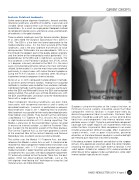
Three anatomic landmarks exist for femoral rotation. Berger
et al. described the Surgical Epicondylar Axis (SEA) in
1993. The SEA is a line from the lateral epicondyle to the
medial collateral sulcus. It is the most accurate of the three
landmarks, and is the only landmark that remains of value
during revision. Whiteside’s line was described in 1995 as a
“line through the deepest part of the patella groove anteriorly
and the centre of the intercondylar notch posteriorly”. It is
perpendicular to the SEA and is slightly less accurate. The
final landmark is the Posterior Condylar Axis (PCA), which
is 3 degrees internally rotated to the SEA. It is the most
easily instrumented and hence remains the most commonly
utilized. Unfortunately it is also the most inaccurate landmark,
particularly in valgus knees with lateral femoral hypoplasia.
Using the PCA in isolation is ill-advised, often resulting in
inadvertent femoral component internal rotation.
Siston et al. in 2005 compared multiple different methods
to achieve correct femoral rotation, finding that navigated
methods in isolation were no better than anatomic methods.
Combined methods had the greatest accuracy, particularly
either the SEA and Whiteside’s line or the SEA and navigated
patella tracking. The author uses all three rotational axis, with
the SEA and Whiteside’s Line drawn onto the resected distal
femur with a surgical marker.
Tibial Component rotational landmarks are even more
03
inaccurate, with no general consensus and a variety of
described landmarks. Lawrie et al. performed a cadaveric Surgeons using minimization of the surgical incision, or
minimally invasive surgery, should be aware that tibial
tibial landmarks study in 2011, reporting that no anatomic
tibial landmark was parallel to their newly described dynamic component internal rotation is made more likely by
diminishing proximal tibial visualization. Posterior tibial
Knee Motion Axis (KMA). They found that the commonly
used Medial 1/3 Tubercle to PCL (Insall’s) Axis resulted retractors should be used with care, as they tend to drive
in slight external rotation of 3.5% during squatting. The tibial trials and components into internal rotation when
posterior access is tight. Trying to maximize tibial component
Medial – Lateral Axis of the resected tibial surface, resulted
in excessive internal rotation, as did the Posterior Tibia coverage with larger implants will inadvertently internally
rotate the tibia. Asymmetric tibial components can make
Axis (tangent to the posterior aspect of the tibial plateau).
The Central Tubercle Axis (a line from geometric centre rotational placement difficult to judge. Most inadvertent
surgical events, such as poor visualization, posterior
of the resected tibia to the central tubercle) and Femoral
Epicondylar Axis resulted in excessive internal rotation.
retractors, or the lateral femoral condyle in a contracted tight
joint tend to push tibial trials into internal rotation.
The author uses the Medial 1/3 Tubercle Axis, but does
If the above anatomic landmarks are indistinct or the surgeon
reference the Medial – Lateral Axis as well. It should also be
realized that tibial component medio-lateral translation also remains intra-operatively uncertain about the correct rotation,
choosing the position with slight external rotation is the
effects rotation, with 1 – 1.5o external rotation per mm of
medial translation. Deeper resection and severe varus makes safest option. As Bell et al. noted in 2012, external rotation of
all landmarks less accurate, with tibial component placement either tibia or femur appears to be asymptomatic, while slight
internal rotation typically results in pain.
10o externally rotated to the Posterior Tibial Axis the best
option. Other non-tibial landmark methods, such as the
“self-aligning – free floating trial” technique or using the 2nd
metatarsal axis are more error prone.
ISAKOS NEWSLETTER 2014: Volume II 25