Page 28 - Layout 1
P. 28
CURRENT CONCEPTS
TKR Component Malrotation: A Common
Unrecognized Cause of Pain & Stiffness
Investigation and Management
The most important diagnostic tool is a high degree of
suspicion and a CT scanning protocol when confronted
with a stiff, painful or dissatisfied TKR. Patients will have
been painful and stiff since implantation, differing from other
common causes of pain such as loosening. Multiple prior
manipulations and second or third opinions will be typical.
Often diagnosis such as Chronic Regional Pain Syndrome,
Arthrofibrosis or metal sensitivity will have been made
previously.
Other causes of pain and stiffness always require exclusion,
such as infection, loosening or referred pain.
05
Malrotated patients often have no clinical signs, but a
reduced range of motion is common. With the patient
Revision should be only undertaken if the patient’s
seated and the knee at ninety degrees, hanging over the bed dissatisfaction warrants a major surgical procedure and its
edge, greater external rotation of the foot on the affected
side can be evident. Patella instability or mal-tracking may associated risks. Patients must be aware that revision may
not result in total satisfaction. Revision of both components
also be present. Plain radiographs typically appear normal,
however, in cases with severe tibial internal rotation, the is usually recommended as both are usually mal-rotated, but
a case can be made for isolated tibial or femoral component
fibular head will appear more covered by the tibia. In Figure revision if the remaining component is aligned, appropriately
3, plain radiographs of a TKR with internal rotation of both
sized and stable. It is vital not to compound the errors of
components reveal subtle increased fibula head coverage. the original procedure. Patella resurfacing or stabilization
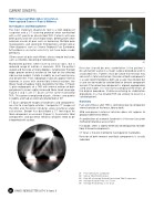
Figure 4 and 5 are CT Scans of the same malrotated
procedures in a mal-rotated TKR are unlikely to result in a
TKR. This patient complained of pain, stiffness and patella satisfied patient.
instability, and had an externally rotated foot at 90o.
CT Scan composite images of landmarks and components Summary
Pain and stiffness after TKR is commonly due to component
are vital to investigate rotation. Composite CT images of
the SEA and Posterior Condyles allow rotational angle internal rotation of the femur, tibia or both.
calculation. Berger has described a CT technique for
Mild component external rotation does not appear to
tibial component assessment (Figure 5). Asymmetric tibial produce detrimental effects.
components and posterior femoral condyles need to be
A combination of anatomic landmarks is the most accurate
interpreted with caution.
method of rotational assessment.
If in doubt, select a slightly externally rotated position for both
tibial & femoral components.
CT Scan is the only method to investigate for malrotation.
Revision of both femoral and tibial components is usually
indicated.
04
01 Femoral Anatomic Landmarks
02 Various Tibial Rotational Axes
03 Plain XR of Internally Rotated Femoral and Tibial Components
04 Internally Rotated Femur on CT Scan
05 Internally Rotated Tibia on CT Scan
26 ISAKOS NEWSLETTER 2014: Volume II