Page 35 - Layout 1
P. 35
CURRENT CONCEPTS
The scope of treatment for hip arthroscopy Although several recent systematic reviews and
continues to expand with many novel techniques metaanalyses have increased the strength of
introduced in the KSSTA special issue. In case evidence-based research, there is still a paucity
reports, the hip arthroscope has been shown of randomized controlled trials in hip arthroscopy
to be valuable with arthroscopic reduction and surgery. Through future research we therefore need
internal fixation of selected acetabular fractures to develop evidence-based guidelines that inform
and femoral head fractures. Hip arthroscopy to surgeons about surgical indications and outcomes
address intra-articular pathology has also been of hip arthroscopy. Furthermore, there is a need to
shown effective as a combined approach with provide results from well-designed investigations
periacetabular osteotomy for dysplastic hips. In addressing areas such as diagnostic algorithms
addition, arthroscopic treatment can be helpful in and surgical outcomes. An ideal course to advance
treating acute septic arthritis of the hip joint in adults hip arthroscopy research would be to gather
and recurrent acetabular osteoid osteoma. Lastly, information about the latest surgical techniques from
Safran et al. have demonstrated an endoscopic expert clinicians, obtain methodological guidance
technique to address ischiofemoral impingement in from researchers, and obtain validated outcomes
the hip by increasing the space between the ischium measures from patients.
and femur.
As the knowledge and understanding of hip disease
While indications continue to expand, complications and arthroscopy continues to grow, we hope this
are not absent from hip arthroscopy. Dietrich ISAKOS newsletter article serves a summary of
et al. emphasize the steep learning curve in hip current literature and stimulates further investigation
arthroscopy and demonstrate the importance into hip arthroscopy with the goal of improving
of training with an expert surgeon. Zingg and outcomes for athletes and patients with hip
colleagues report a 1.9% rate of femoral neck conditions. Ultimately, high-level research will be
insufficiency fractures following cam resection crucial for improving hip arthroscopy as a successful
and femoral neck osteochondroplasty, and offer treatment for patients with disorders of the hip.
guidelines for postoperative rehabilitation. On the
soft tissue side, Smith et al. demonstrate that
the capsular restraints in the hip are essential to
maintaining normal hip biomechanics. In revision hip
arthroscopy, McCormick et al. report that capsular
defects are relatively common, which may lead to
worse outcomes. In addition to capsular defects,
Willimon et al. show that adhesions can develop
following hip arthroscopy, with a higher likelihood
in patients less than 30 and rehabilitation without
circumduction.
As the field of hip arthroscopy and hip preservation
is still young, there is ample opportunity for future
research. Surgeons and scientists, who critically
evaluate scientific literature must review and refine
indications and surgical techniques. Similarly, we
must improve our understanding of rehabilitation
procedures and for measures of (re-) injury
prevention. Future research is required for the 05
enhancement of surgical repair procedures with
biologics, such as growth factor delivery, stem
cells, or tissue engineered materials. Rigorous
future research, critical appraisal of indications and
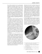
01 Intraoperative arthroscopy photo demonstrating exposure of
techniques, as well as peer-review will then assure a large cam lesion through a T-cut capsulotomy.
advancing of hip arthroscopy and ultimately help
02 Intraoperative arthroscopy photo demonstrating cam lesion
improving outcome for our patients.
resection and recontouring of the femoral neck through a
T-cut capsulotomy.
03 Intraoperative arthroscopy photo demonstrating closure of
the T-cut capsulotomy.
04 Preoperative frog leg lateral view of distal cam bump with
loss of femoral neck offset.
05 Postoperative frog leg lateral view of cam bump resection
and femoral neck recontouring.
ISAKOS NEWSLETTER 2014: Volume II 33