Page 36 - Layout 1
P. 36
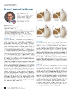
Freehill Fig 1


CURRENT CONCEPTS


Bennett’s Lesion of the Shoulder

AB


Michael T. Freehill, MD
Department of Orthopaedic Surgery,

Division of Sports Medicine and
Shoulder Surgery, Wake Forest
University School of Medicine,

Medical Center Boulevard, Posteroinferior subperiosteal lesion
Posteroinferior free fragment
Winston-Salem, NC, USA


Additional Authors:
CD
Sandeep Mannava, MD, PhD1
Laurence D. Higgins, MD2

1 Department of Orthopaedic Surgery, Division of Sports Medicine

and Shoulder Surgery, Wake Forest University School of Medicine,
Medical Center Boulevard, Winston-Salem, NC, USA
2 Department of Orthopaedic Surgery, Sports Medicine and

Shoulder Service, Brigham and Women’s, Harvard University
School of Medicine, 75 Francis Street, Boston, MA, USA
Posterosuperior Posterosuperior
subperiosteal lesion
subperiosteal free
Introduction
fragment
01
Throwers’ exostosis (spurring or bony formation) is a
calcification arising in the posterior region of the glenoid. The Presentation

lesion was first described in professional baseball pitchers Bennett lesions are often associated with undersurface
by George E. Bennett in 1941, who stated it was one of
rotator cuff tears and posterior labral injury. Meister et al.
the distinctive lesions of the shoulder which could end the reported 95% (21/22) undersurface cuff tears and 68%
career of the professional pitcher. More recently, Wright and
(15/22) with posterior labral pathology in throwers with
Paletta reported the prevalence of such lesions at 22% in Bennett’s lesions. Both of these associations are likely
55 asymptomatic major league pitchers. Despite awareness secondary to posterior internal impingement, with the
of its presence in throwers, the most effective management
Bennett lesion formation on the posterior glenoid. Therefore,
and technical considerations of surgery are still debatable. determination of whether the pain generation is secondary to
Returning elite overhead athletes to symptom free throwing
the Bennett lesion or the associated pathology, is a critical
is a difficult proposition. This is evidenced by a lack of component of evaluation and treatment.
consensus in management of the condition and underscored
Based upon a comprehensive review of the literature,
by a paultry 55% rate of return to successful pre-injury levels
of throwing in the hands of skilled surgeons familiar with increased pain appears associated with a Bennett free
fragment, whereas glenohumeral internal rotation deficit
caring for elite baseball players.
(GIRD) appears more prevalent in the setting of an attached
Interestingly, numerous lesions have been described under
lesion.
the title of Bennett lesion, yet they differ in presentation.
Diagnosis
Bennett described the classic eponymous lesion, located
in the posteroinferior region of the glenoid. This exostosis
It has been demonstrated that the occurrence of Bennett
has been reported to be subperiosteal attached to the
lesions, both painful and asymptomatic, increased
glenoid and as a free bony fragment. Subperiosteal lesions
significantly with advanced age and duration of throwing.
have similarly been reported at the triceps attachment. The
Yoneda et al. described criteria for diagnosing a painful
“superior Bennett lesion” was described by Nakagawa et al. Bennett lesion which included: detection of the spurring
and differs from a conventional Bennett lesion in location, as or lesion on the posterior rim of the glenoid, presence

it is occurring in the region of the posterosuperior glenoid rim. of posterior shoulder pain with throwing, tenderness to
Again, the “superior Bennett lesion” can be subperiosteally palpation at the posteroinferior aspect of the glenohumeral

attached to the glenoid or as a free bony fragment. Finally, joint, and improvement of throwing pain following injection
the “pitcher’s mound” osteophyte was described by Pearce of xylocaine around the lesion. Therefore, in the overhead

and Burkhart at the posterosuperior glenoid rim associated thrower with complaints of posterior shoulder pain, the
with type II SLAP tears. (Figure 1A-D)
existence of a painful Bennett lesion should be considered.

A Bennett lesion can be a subtle finding on plain film

radiography, but specific views have been reported effective
to increase identification of the lesion. (Figure 2)


34 ISAKOS NEWSLETTER 2014: Volume II